Quarter horse
Male castrated
9 years of age
Chronic lameness hind right: Anamnesis of trauma 3 years before
Since then intermittently lame, for short time severe lameness and then better again
Treated for osteoarthritis in the tarsometatarsal joint
Complete study of the right tarsus was performed
Radiographic examination
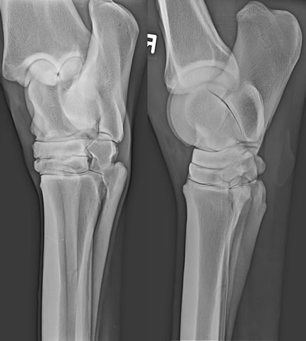
Dorsolateral-plantaromedial, lateromedial, dorsoplantar and dorsomedial-plantarolateral view of the right tarsus.
Radiographic findings and diagnosis
- There is irregularly defined tarsometatarsal joint, subjectively narrowed and surrounded by bone sclerosis.
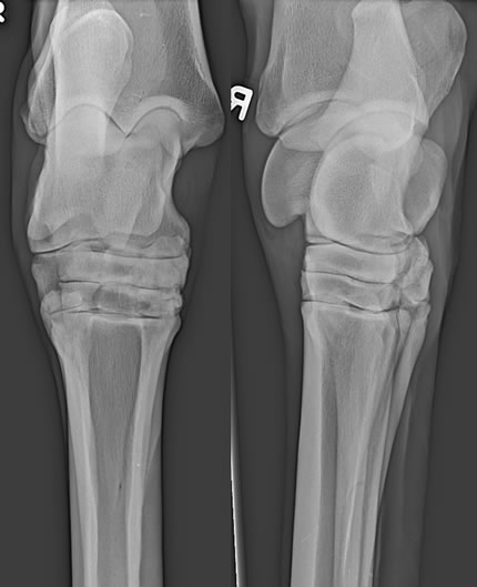
- 2 projections of the left tarsus were also taken for comparison.
Radiographic examination
Dorsomedial-plantarolateral and lateromedial view of the left tarsus.
- The left distal intertarsal joint was read as normal.
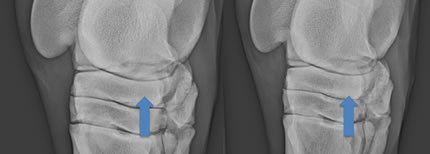
- Comparing the same 2 views of the tarsi, there is a severe sclerosis of the central tarsal bone on the right side (arrow) with focal loss of the trabecular structure.
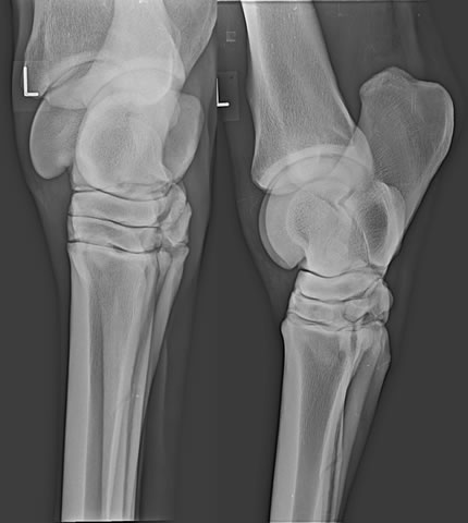
Radiographic examination
Close up of the dorsomedial-plantarolateral view of the tarsi (left on the left image, right on the right image)
The horse underwent CT examination.
Radiographic examination
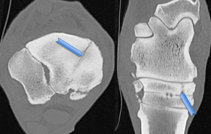
CT, bone algorithm, transverse view (image on the left) and multiplanar reconstruction (on the right), dorsal plane.
- There is a biarticular fracture of the central tarsal bone (arrowhead), running from dorsomedially to plantarosagittal.
- The fracture is surrounded by marked bone sclerosis.
Comments
- The radiographic diagnosis was fracture of the right central tarsal bone, biarticular and not dislocated.
- Also retrospectively, the fracture on the radiographic study could not be identified.
- The sclerotic appearance of the central tarsal bone, appreciated on the radiographs, was interpreted secondary to the osteoarthrosis.
- Most central tarsal fractures are complete, breaking into both the proximal and distal intertarsal joints. Some authors theorize that such injuries are the result of accumulated bone damage, so-called microtrauma, which eventually leads to overt structural failure.
- Fractures of the central tarsal bones are difficult to detect with radiographs because of the complex anatomy of the tarsus and the superimpositions of the tarsal bones. Often several oblique views are needed to identify the fracture line. Sometimes, in a later re-check, 7-10 days after the fracture, rarefaction of the fracture margins will make the fracture line more obvious.
- The fracture was fixed as shown in the post OP radiographs.
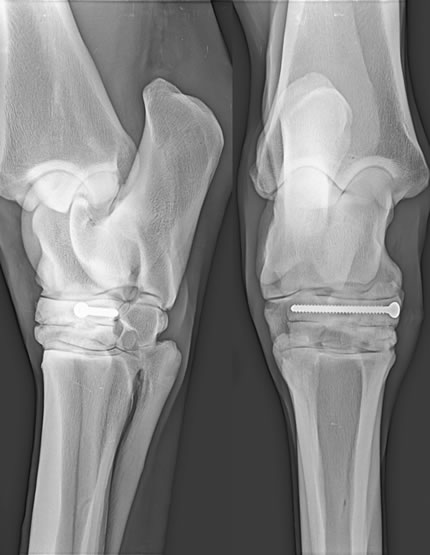
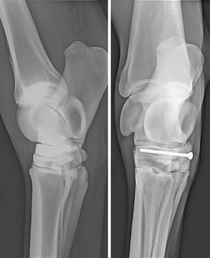
Dorsolateral-plantaromedial, lateromedial, dorsoplantar and dorsomedial-plantarolateral view of the right tarsus post OP.


